|
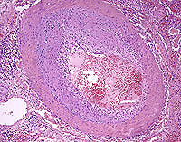
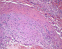
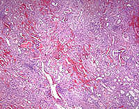
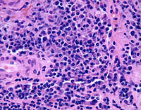
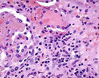
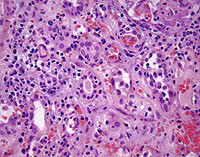
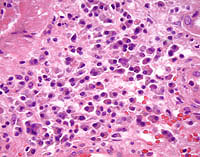
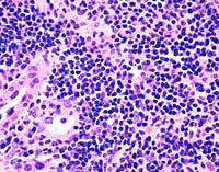
Case Control Study of Severe PTLD in Renal Transplant Recipients. Case 1. Severe kidney rejection with plasmacellular features. This renal transplant patient had an echogenic mass located in the hilum of the allograft kidney. The mass was considered to be consistent with a PTLD, and it caused renal vascular stenosis and hydronephrosis. The photomicrographs are from the allograft nephrectomy which was performed 6 days later. Both acute and chronic rejection were present in this specimen. Lymphoplasmacytic aggregates were initially considered to be suspicious for, but not diagnostic of, PTLD. A followup immunocytochemical stain for EBV was performed and this was negative. No evidence of PTLD was seen in this specimen, and this was stated in the final diagnosis. The histologic review is in agreement with this interpretation.
(Please note: Forward button takes you to photomicrographs of Case 1. When additional cases are added, it will be changed to take you to the next case) Please mail comments, corrections or suggestions to the TPIS administration at the UPMC.
If you have questions, please email TPIS Administration. |
||||