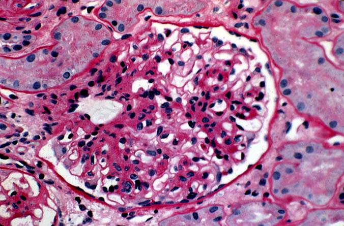
Histopathological examination in recurrent IgA nephropathy most commonly
shows increased mesangial matrix or mild mesangial proliferation.
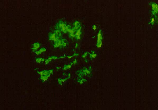
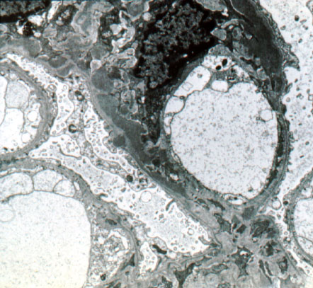
Immunofluorescence examination and electron microscopy confirm the presence
of IgA immune complex deposits in a mesangial location. Other patterns of
glomerular injury include minimal change, endocapillary proliferation,
membranous nephropathy and combined lesions.
Please mail comments, corrections or suggestions to the
TPIS
administration at the UPMC.