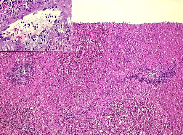
Figure 1.
This example of severe acute rejection was taken from a failed allograft,
removed several weeks after transplantation.
Note the marked portal tract(PT) inflammation that focally spills
over into the periportal hepatic parenchyma. A similar infiltrate is
seen in and around the
central veins(CV), and is associated with perivenular hepatocyte
necrosis and dropout.
The inset(upper left) shows an inflammatory and necrotizing arteritis
with early focal foam
cell deposition. This artery was present in a section taken from the
hilum of the failed allograft.
Arteritis is rarely detected in needle biopsies because the vessels
most commonly affected
are contained within the liver hilum and rarely sampled with a needle biopsy.
Please mail comments, corrections or suggestions to the
TPIS administration at the UPMC.