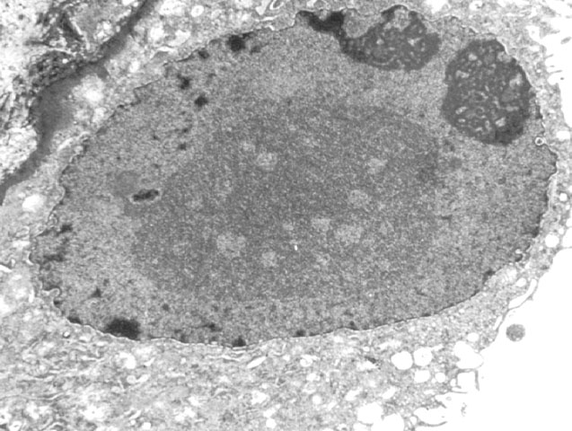
Figure 3.
Electron micrograph prepared from a biopsy with BK virus infection
showing a tubular epithelial cell packed with virions. The nuclear
chromatin shows peripheral margination.
Please mail comments, corrections or suggestions to the
TPIS
administration at the UPMC.