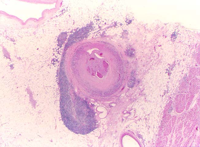
Figure 4.
This low power photomicrograph illustrates GVD involving an epicardial coronary
artery. Note the concentric intimal thickening, the adventitial inflammation
and the superimposed lumenal thrombosis, which is showing partial
recanalization.
Please mail comments, corrections or suggestions to the
TPIS
administration at the UPMC.