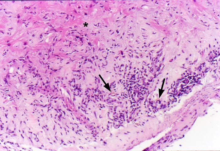
Figure 6.
A higher power of the photomicrograph in Figure 5, illustrates the myofiber
disarray(*), fibrosis, sparse mononuclear inflammation and pigmented
macrophages(arrows),
typical of previous biopsy sites, which have largely healed.
Please mail comments, corrections or suggestions to the
TPIS
administration at the UPMC.