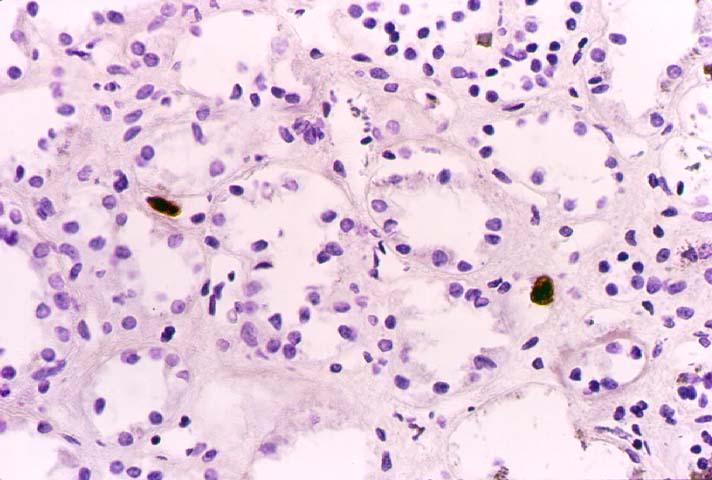
Figure 3.
Immunoperoxidase staining highlighting the presence of
cytomegalovirus infected cells with marked nuclear
enlargement. It is difficult to be certain whether the
cells shown belong to the peritubular capillary
endothelium or represent atrophic tubular epithelium.
Please mail comments, corrections or suggestions to the
TPIS
administration at the UPMC.