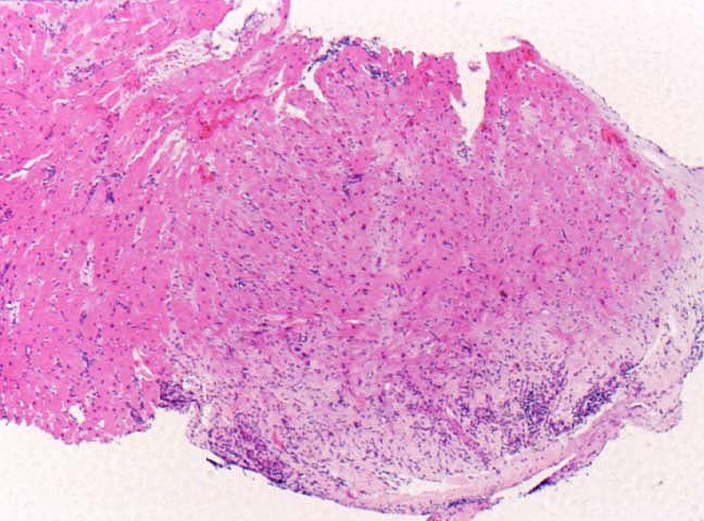
Figure 5.
Previous biopsy sites are seen in up to 30% or more
of endomyocardial biopsies obtained within the first 30 days
after transplantation. If they are sampled soon after the
previous biopsy, partially organized fibrin masses are usually
detected. However, if they are sampled more than several
months after the previous biopsy, there may only be a focal area
of fibrosis, accompanied by pigmented macrophages, plasma
cells and occasional lymphocytes. The key identifying feature
however, is the presence of myofiber disarray at the base of the
lesion(arrow).
Please mail comments, corrections or suggestions to the
TPIS
administration at the UPMC.