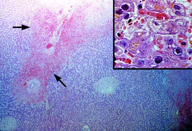
Figure 5.
Occasionally, the diagnosis of herpes simplex or varicella-zoster
virus infection can be made directly from the routine stains. This low power
photomicrograph of a failed liver allograft was obtained 3 days after hepatic
transplantation. Note the large areas of coagulative-type necrosis scattered
randomly throughout the sample [arrows]. The inset shows the Cowdry type A
inclusions located in the viable cells at the edge of the necrotic zones.
Please mail comments, corrections or suggestions to the
TPIS administration at the UPMC.