Figure 1.
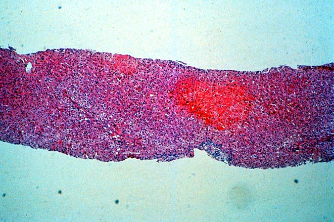
This low power photomicrograph shows the typical appearance of
herpes simplex or varicella-zoster viral hepatitis in a liver allograft.
Note
the punched-out areas of coagulative-type necrosis scattered randomly
throughout
the biopsy without respect for the architectural landmarks.
Please mail comments, corrections or suggestions to the
TPIS administration at the UPMC.