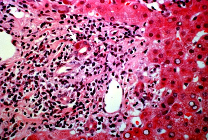
Figure 9.
This is a higher magnification of the portal tract and surrounding
periportal hepatic parenchyma. Note the cytomegalovirus inclusion body present
in the enlarged biliary epithelial cell [arrow]. Note also the
lymphoplasmacytic infiltrate present in the portal tract, which can mimic acute
cellular rejection if one misses the inclusion bodies.
Please mail comments, corrections or suggestions to the
TPIS administration at the UPMC.