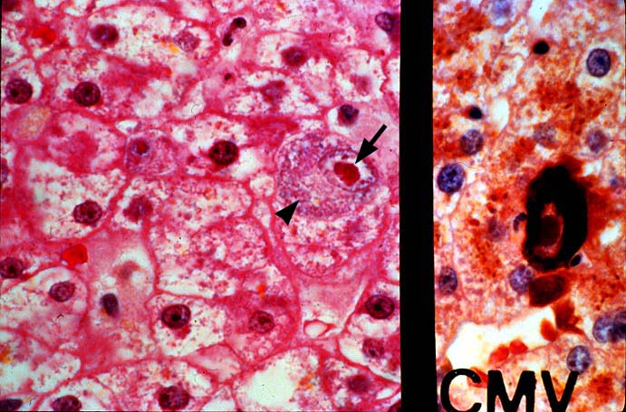
Figure 7.
This high power photomicrograph shows the characteristic nuclear and
cytoplasmic inclusions typically seen with cytomegalovirus. In the left side
of the photomicrograph note the enlarged or cytomegalic cell containing both
nuclear and cytoplasmic inclusions. The cytoplasmic inclusions are usually
small and basophilic(arrowhead). The nuclear inclusions are usually much
larger, eosinophilic and show a halo or clear space between the inclusion in the
nuclear membrane(arrow). The right side of the photomicrograph shows a similarly
infected cell stained for early and made cytomegalovirus antigens.
Please mail comments, corrections or suggestions to the
TPIS administration at the UPMC.