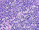
Neoplastic PTLD. Case 2.
Polymorphic diffuse B cell hyperplasia in a renal transplant recipient
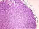
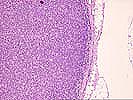
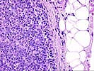
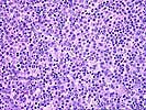
This young boy developed a PTLD 10 months following liver transplantation. This example is from an involved lymph node. The diffuse infiltrate effaces most of the nodal architecture except for a few residual sinusoids. The infiltrate does not appreciably transgress the capsule. A variety of mononuclear cells are seen, with a low power impression of a dimorphic large cell and small lymphocyte population. On closer examination, the large cells consist of significant numbers of immunoblasts, with lesser numbers of centroblasts and scattered centrocytoid cells or centrocytes. Some plasmacellular features are noted. Occasional immunoblasts contain rectangular nucleoli and could be of B or T cell origin. Atypical features such as irregular chromatin dispersal or convoluted nuclear membranes or multilobation are not seen. The evenly dispersed smaller cells consist of small lymphocytes and small cleaved cells (centrocytes). Scattered individual apoptotic cells are seen, as are scattered mitoses. The overall features are most compatible with the diagnosis of polymorphic diffuse B cell hyperplasia. In Pittsburgh nomenclature, the lesion would be considered a polymorphic PTLD. Immunoglobulin gene rearrangement analysis showed a monoclonal B cell population in this tumor. Extranodal disease was also present in this patient. He was treated conservatively and the lesions resolved. As of last followup, the patient was alive 5 ½ years after the diagnosis of PTLD.
|
|
|
|
Note: Click on the thumbnails to view the microscopy of this case- the "up" arrow will take you to the top of the page to see them again. Click on the forward button to go to the next case. Click on the back arrow to return to the previous case.