Previous Biopsies on this Patient:
None
TPIS Related Resources:
Liver Allograft Rejection Grading
Liver Transplant Topics
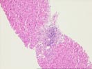
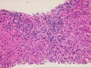
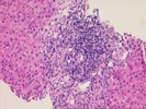
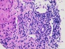
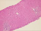
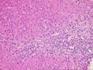
(Part 1: 2 HE, 1 Trichrome, 1 PAS/D)
The normal lobular architecture is distorted by mild to moderate
portal expansion because of fibrosis and a moderate mononuclear
inflammatory cell infiltrate. Duct damage is clearly appreciated
in half or more of the triads. Focal duct loss may be developing
in approximately four of ten triads. There is also mild
interface activity.
Throughout the lobules, there is Kupffer cell hypertrophy and occasional acidophilic bodies. Occasional foam cell clusters are also seen. However, the most striking change is the prominent central venulitis with perivenular hepatocellular dropout. No definite viral inclusions or ground glass cells are seen. The PAS/D stain nicely shows the bile duct infiltration and damage, with lymphocytes located inside the basement membrane.
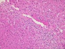
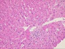
(Part 2: 2 HE, 1 PAS, 1 PAS/D, 1 Iron, 1 Reticulin) The normal lobular architecture is slightly distorted secondary to mild to moderate portal expansion because of mild fibrosis and a mild, predominantly mononuclear inflammatory cell infiltrate. Focal bile duct infiltration and damage are easily seen.
Throughout the lobules, there is Kupffer cell hypertrophy and occasional acidophilic bodies. However, there is little evidence of disarray. The most striking lobular change is the presence of centrilobular hepatocellular inflammation, dropout and hemorrhage. This is associated with a perivenular mononuclear inflammatory cell infiltrate containing lymphocytes, plasma cells and pigmented macrophages. No definite viral inclusions or ground glass cells are seen.
Overall, the histopathologic changes in both biopsies are most consistent with an active cellular rejection with prominent central venulitis. Bile duct damage and focal bile duct loss are also easily seen.