Previous Biopsies on this Patient:
None
TPIS Related Resources:
Liver Allograft Rejection Grading
Liver Transplant Topics
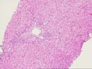
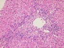
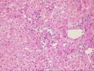
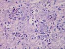
| 1. Histological Evaluation | ||||
|---|---|---|---|---|
| 1.1 More than 4 portal triads | (X)YES | ( )NO | ||
| 1.2 Specimen o/w adequate | (X)YES | ( )NO | ||
| 2. Portal Tract (check one uner each category) | ||||
| 2.1 Overall Inflammatory Intensity | ( )None | (X)Mild | ( )Moderate | ( )Severe |
| 2.2 Bile duct inflammation / damage | (X)YES | ( )NO | ||
| 2.3 Granuloma(s) | ( )YES | (X)NO | ||
| 2.4 Bile duct loss | ( )YES | (X)NO | ||
| 2.4.1 If BD loss, total number of triads | ( ) | |||
| 2.4.2 If BD loss, number of triads w/o ducts | ( ) | |||
| 2.5 Bile duct / cholangiolar proliferation (in any portal tract) | (X)YES | ( )NO | ||
| 3. Inflammatory or Necrotizing Arteritis | ( )YES | (X)NO | ||
| 4. Obliterative Arteriopathy | ( )YES | (X)NO | ||
| 5. Subendothelial Inflammation | ||||
| 5.1 Severity | ( )None | (X)Mild | ( )Moderate | ( )Severe |
| 5.2 Location | ( )N/A | ( )Portal | ( )Central | (X)Both |
| 6. Fibrosis (check one under each category) | ||||
| 6.1 Portal | ( )None | (X)Mild | ( )Moderate | ( )Severe (bridging) |
| 6.2 Central | ( )None | (X)Mild | ( )Moderate | ( )Severe (bridging) |
| 6.3 Arch. dist. | (X)None | ( )Mild | ( )Moderate | ( )Severe |
| 7. Lobular Disarray / Ballooning | ( )None | (X)Mild | ( )Moderate | ( )Severe |
| 8. Necrosis (check one under each category) | ||||
| 8.1 Piecemeal or bridging | ( )None | (X)Mild | ( )Moderate | ( )Severe |
| 8.2 Infarct (ischemia) | ( )YES | (X)No | ||
| 8.3 Other necrosis | (X)YES | ( )No | ||
| 8.4 Central lobular | (X)YES | ( )No | ||
| 9. Cholestasis | ( )YES | (X)NO | ||
| 10. Fat | (X)None | ( )Mild | ( )Moderate | ( )Severe |
| 10.1 Type | ( )Micro | ( )Macro | ( )Mixed | (X)N/A |
| 11. Lobular Inflammation (check one under each category) | ||||
| 11.1 Severity | ( )None | ( )Mild | (X)Moderate | ( )Severe |
| 11.2 Location | ( )N/A | ( )Perivenular | ( )Panlobular | (X)Random / Focal |
| 11.3 Granulomatous | ( )YES | (X)NO | ||
| 12. Rejection Activity Index (RAI) | Range | Score | ||
| 12.1 Portal inflammation | (0-3) | (1) | ||
| 12.2 Bile duct inflammation / damage | (0-3) | (1) | ||
| 12.3 Venous endothelial inflammation | (0-3) | (1) | ||
| 12.4 Total Score | (RAI = 3/9) | |||
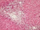
Scattered single Councilman-type bodies are seen. Viral inclusions are not appreciated on routine stain. Focal spotty inflammation is also noted. Portal inflammation shows minimal involvement of portal structures with prominent ductular proliferation at sites. Some piecemeal necrosis is seen, evidenced as single Councilman bodies at the limiting plate area.