Previous Biopsies on this Patient:
NONE
TPIS Related Resources:
Liver
Allograft Rejection Grading
Liver
Transplant Topics


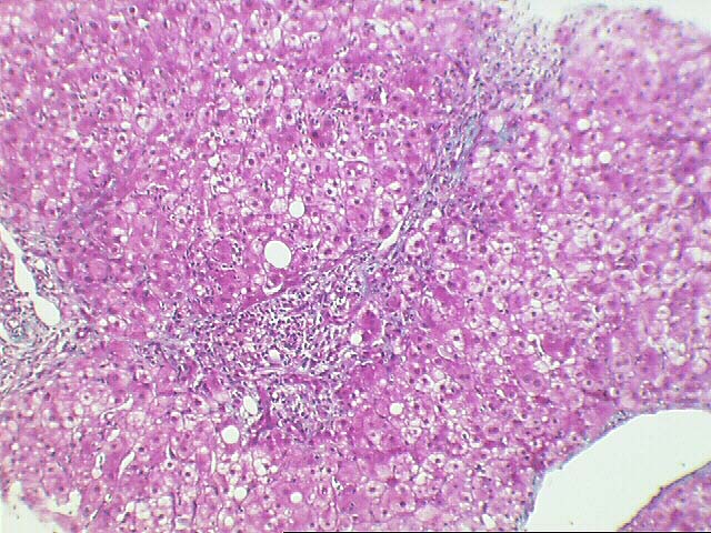
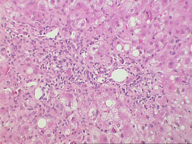
The normal lobular architecture is distorted by moderate portal expansion because of duct and cholangiolar proliferation, mild mixed inflammation and mild portal fibrosis. Very focal and thin band of portal to portal and portal to central bridging fibrosis is identified.
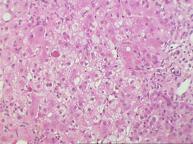
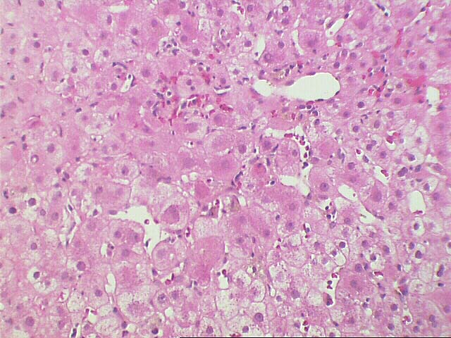
On closer examination, there is no evidence of subendothelial infiltration of either the portal or central veins. No lymphocytic ductulocentric inflammation is appreciated. Instead, activity is prominent at the interface zone where the cholangiolar reactivity and lymphoplasmacytic infiltrate extend into the periportal parenchyma.
Throughout the lobules there is moderate to severe lobular disarray, mild macrovesicular steatosis, Kupffer cell hypertrophy, regenerative change, and moderate to marked spotty acidophilic necrosis of hepatocytes. No definite ground glass cells or viral inclusions are seen.
Overall, the histopathologic changes are indicative of a moderate to severely aggressive hepatitis, consistent with the clinical history of viral type C. This contention is based on the marked lobular and interface activity instead of the vascular and ductuolcentric inflammation seen with acute rejection. Moreover, the marked cholangiolar proliferation, hepatocellular swelling and degeneration, are reminiscent of changes seen with the fibrosing cholestatic variant of hepatitis which can be seen with both hepatitis B and C virus infection. There is little to no evidence of active cellular rejection. Application of the Knodell scoring index is as follows:
| KNODELL'S HISTOLOGY ACTIVITY INDEX | ||
|---|---|---|
| Feature | RANGE | SCORE |
| Periportal and bridging necrosis | (0-10) | 4 |
| Intralobular degeneration and necrosis | (0-4) | 3 |
| Portal inflammation | (0-4) | 3 |
| Fibrosis | (0-4) | 1-3 |
| TOTAL | (0-22) | 11-13 |
The numerical scoring system of histological activity index (HAI) has been developed to grade the liver biopsies of chronic active hepatitis. This is based on four categories of periportal and bridging necrosis, intralobular degeneration and necrosis, portal inflammation and fibrosis, with total score of up to 22. This scoring system is correlated well with the severity of disease. A copy of the original paper published by American Association for the Study of Liver Disease [Hepatology 1:431 (1981)] is available at the Department of Pathology upon request.