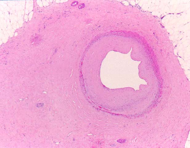
Figure 7.
A higher magnification of one of the arteries shown in figure 6, illustrates the dense adventitial fibrosis of an artery with the later stages of GVD.
Please mail comments, corrections or suggestions to the
TPIS administration
at the UPMC.
[FRAMES]
[NO FRAMES]