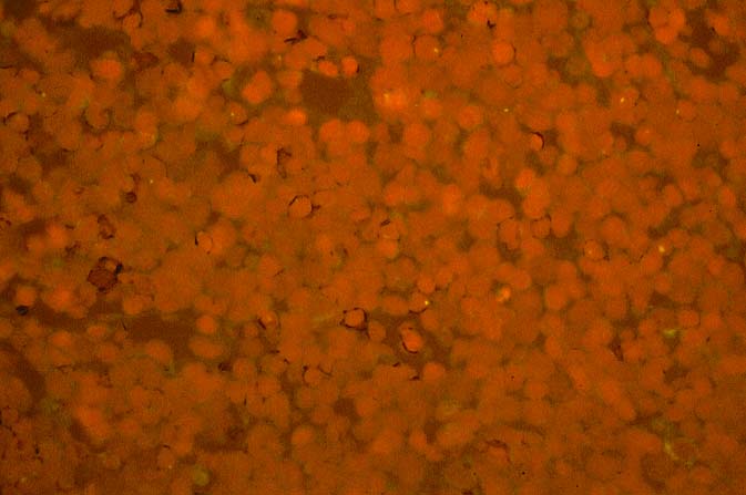
Figure 11.
EBV-related post-lymphoproliferative disorders in the early
post-transplant period not infrequently will contain a significant number of
donor T- lymphocytes, as shown in this photomicrograph, which is simultaneously
stained for T-lymphocytes [CD3; brown stained cells] and for the Y-chromosome
[yellow dots]. This biopsy was taken from an enlarged cervical lymph node of a
47 year old female, who 7 months after transplantation, noted a swelling in her
neck. The liver donor was a 25 year old male. Note the male cells marked with
the yellow nuclear dot, some of which are also T-lymphocytes.
Please mail comments, corrections or suggestions to the
TPIS administration at the UPMC.