Figure 5.
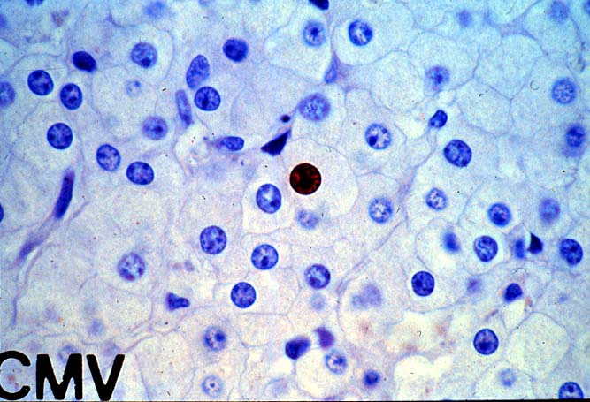
This section was stained for a variety of cytomegalovirus antigens
using a soup of monoclonal antibodies directed against both early and late viral
antigens. Note the positively staining nucleus in the center of the field in a
cell that does not show cytomegalic change.
Please mail comments, corrections or suggestions to the
TPIS administration at the UPMC.