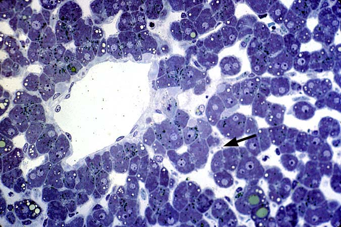
Figure 2.
This plastic embedded section shows evidence of a little more severe
cold preservation injury and the differential sensitivity of the
endothelium lining the larger vessels as opposed to the sinusoids.
Note the intact endothelium lining the central vein in this
photomicrograph. In contrast, many of the sinusoidal endothelial cells
are difficult to recognize, because they are detached from the
underlying connective tissue and have assumed a "rounded" appearance.
Note also the "blebs" on the surface of hepatocytes(arrows). These
are small protrusions on the cytoplasmic membrane of damaged
hepatocytes, and are a marker for hepatocellular injury.
Please mail comments, corrections or suggestions to the
TPIS administration at the UPMC.