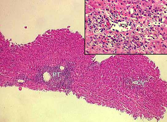
Figure 6.
Severe acute rejection can also be diagnosed in a needle biopsy. In this
photomicrograph,
note the presence of an inflammatory infiltrate expanding the portal
tracts(PT). Note also the
presence of inflammation in and around the central veins(CV). The inset
shows the centrilobular region in greater detail. Lymphocytes are
seen under the endothelium of the
central vein, and extending out into the perivenular hepatic
parenchyma. The associated
hepatocyte necrosis and dropout are typical of a grade "3" venous
damage in the Banff schema.
Please mail comments, corrections or suggestions to the
TPIS administration at the UPMC.