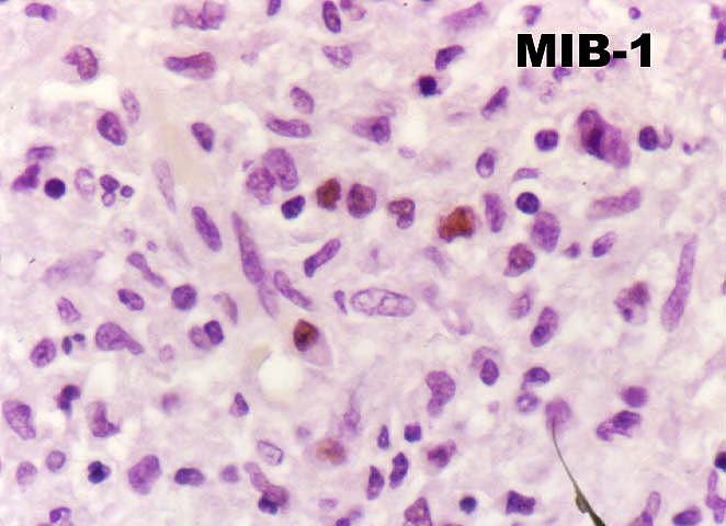
Figure 11.
This is a higher magnification of the shoulder region of the plaque shown
in figure 10 stained with MIB-1, which decorates cells synthesizing
DNA. Note the uptake of cells in the shoulder region, which are a mixture
of donor and recipient lymphocytes. In this case, one
could postulate that an MLR was occurring in the arterial wall.
Please mail comments, corrections or suggestions to the
TPIS
administration at the UPMC.