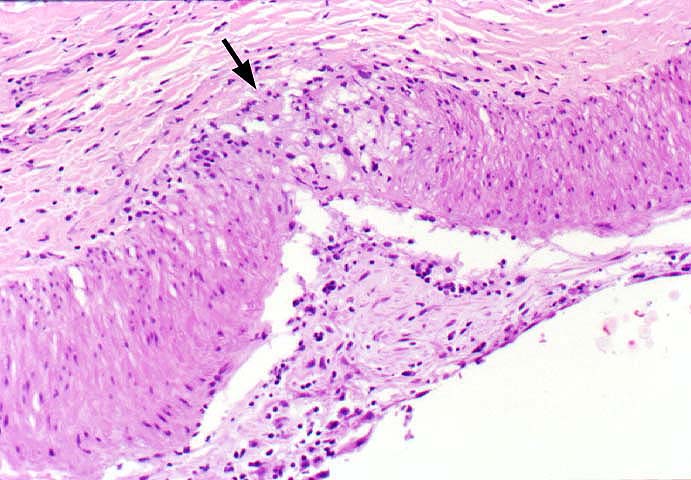
Figure 2.
A higher magnification of the artery shown in figure 1 illustrates the
subintimal foam cell deposition, which focally extends through the media
into the adventitia. Note also the lymphocytes in the thickened intima.
Please mail comments, corrections or suggestions to the
TPIS
administration at the UPMC.