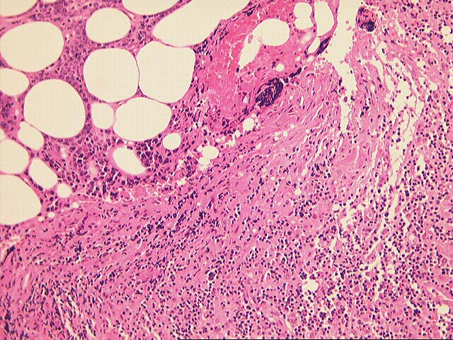
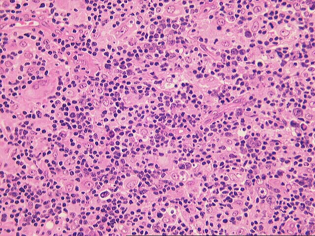
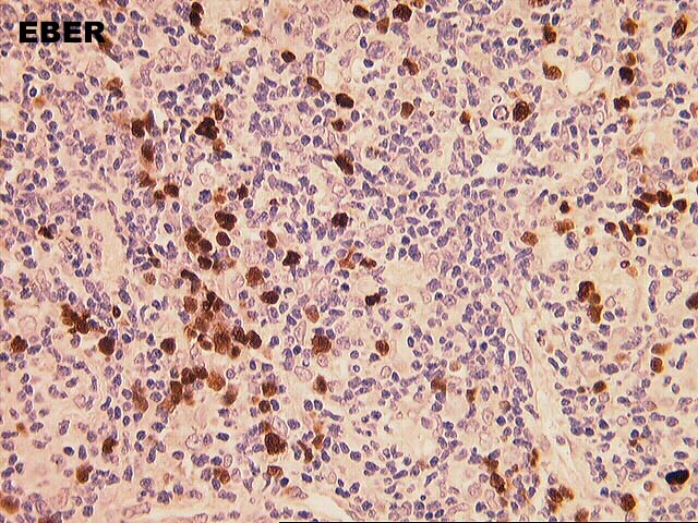
PART 2: MEDIASTINAL LYMPH NODE, LEVEL 9, EXCISIONAL BIOPSY
-
Previous Biopsies on this Patient:
NONE
TPIS Related Resources:
Kidney
Transplant Topics
Neoplasia
Transplant Topics
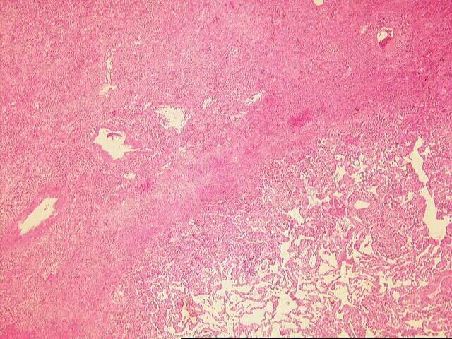
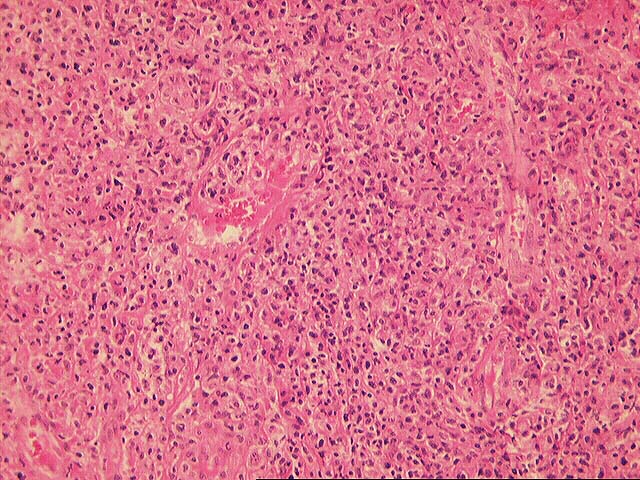
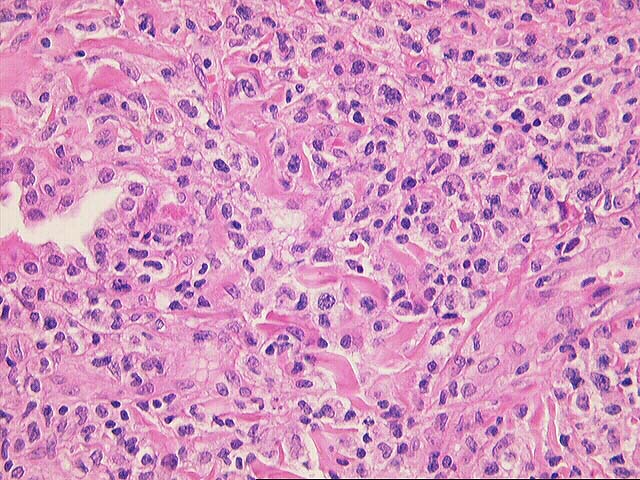
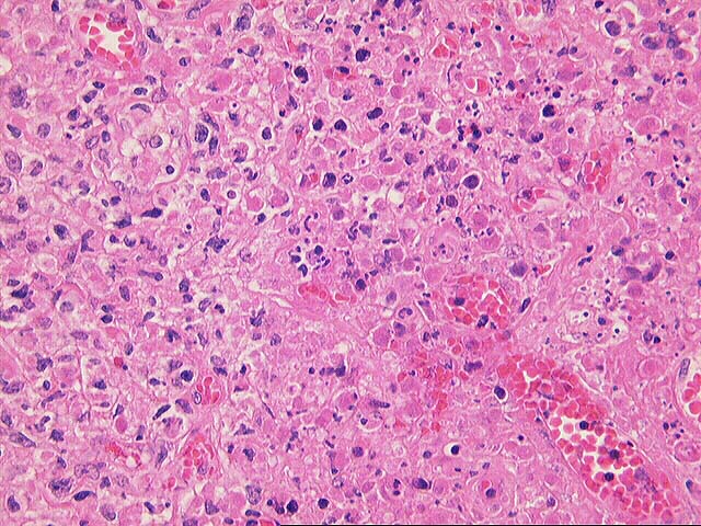
Sections from the lung show the parenchyma partly replaced by a lymphomatous infiltrate comprised of large non-cleaved cells, immunoblasts and small cleaved/non-cleaved cells. Focal areas of necrosis are present, and these are associated with a significant influx of neutrophils. Focal vasculitis is present in the lesion. No Reed-Sternberg cells are seen.
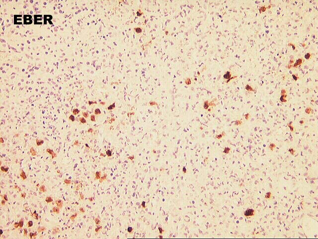
The mediastinal lymph node is largely replaced by a lymphomatous infiltrate similar to that described in the lung biopsy. However, there is less necrosis in this specimen, and some microscopic fields consist predominantly of large cells. EBER stains are strongly positive, and more so than in the lung specimen.
Insufficient unstained slides have been received for immunophenotypic studies, but tissue was sent for flow cytometry and gene rearrangements in the referral hospital.