 |
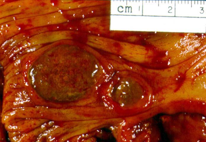
| Figure 1. PTLD involvement of large bowel. Two adjacent but noncontiguous ulceronodular lesions are seen on the luminal aspect of this large bowel specimen. The borders have a slightly heaped-up appearance, but the process subsides almost immediately beyond the confines of the ulcerated masses. The green discoloration corresponds to loss of mucosal lining cells with overlying fibrinoinflammatory debris, often with large numbers of bacteria. |