 Contributed by Anthony J. Demetris, MD and Michael S. Torbenson, MD
PATIENT HISTORY:
Patient with cirrhosis, hepatic lesion and liver failure. The referring pathologist believes this biopsy is suspicious for hepatocellular carcinoma.
Final Diagnosis (Case 101)
PART 1: NATIVE LIVER, NEEDLE BIOPSY -
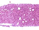
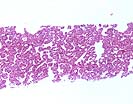
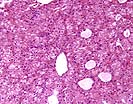
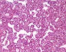
- WELL DIFFERENTIATED HEPATOCELLULAR CARCINOMA
- NO NON-NEOPLASTIC LIVER IDENTIFIED.
Microscopic Description - (Case 101)
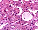
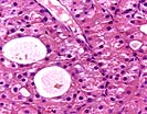
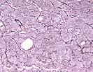
Sections of liver are entirely composed of neoplastic tissue. No normal liver architecture or portal tracts are seen. The neoplastic cells are growing in cords that range from 1 to 6-7 cells thick, and pseudoacinar structures, interrupted by occasional aberrant "feeding" arteries. The neoplastic hepatocytes show abundant pink cytoplasm with generally round nuclei and generally inconspicuous nucleoli, though occasional more prominent nucleoli are seen. Bile production is also clearly seen. A reticulin stain demonstrates preservation of the reticulin framework in most of the tumor, but definite focal loss of reticulin is seen. An iron stain shows mild iron deposition, principally in Kupffer cells. A trichrome stain demonstrates mild and focal pericellular fibrosis. A PAS-D stain is negative for intracytoplasmic globules.
No non-neoplastic liver is present and the extent of background liver disease can not be evaluated. However, by history, it is said to be cirrhotic.
Overall, it is my opinion that the histopathologic changes warrant a diagnosis of well-differentiated hepatocellular carcinoma. This contention is based on the thickness of the plates, nuclear density in areas, the "aberrant feeding" vessels, the focal loss of reticulin framework and the history of cirrhosis.
TPIS Related Resources:
Gross Description - (Case 101)
Received for consultation five (5) consult slides along with an accompanying surgical pathology report.
Photomicrographs - (Case 101)
Please mail comments, corrections or suggestions to the
TPIS administration at the UPMC.


