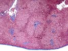
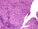
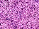
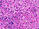
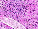
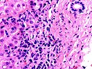
Within the lobules, the sinusoids show a scattered predominately lymphocytic infiltrate and a rare necrotic hepatocyte. Mild and focal cholestasis is also seen both within hepatocytes as well as bile canaculi. Scattered binucleated forms and occasional pseudorosette formation evidence lobular regenerative activity. Scattered hepatocytes also show glycogenated nuclei. The central veins show a minimal increase in fibrous tissue as well as a lymphoplasmacytic infiltrate within the zone 3 hepatocytes, accompanied by occasional hepatocyte drop out and occasional extravasated red blood cells. No granulomas, viral inclusions, nor florid duct lesions are identified. A trichrome stain highlights the mild portal fibrosis. No increased fat deposition or pericellular fibrosis is seen.
Overall, the histopathological findings are most consistent with an active hepatitis. Possibilities would include prolonged hepatitis A viral hepatitis, autoimmune hepatitis and an adverse drug reaction. The first of these possibilities, prolonged hepatitis A, is the favored interpretation. This opinion is based on the well-known association of hepatitis A with a "chronic hepatitis histology"; the prominent plasmacytic component, also reported in hepatitis A; the clinical history of hepatitis A; and the active lobular inflammation with cholestasis that can also be seen with hepatitis A. Re-testing of anti-HAV antibodies for IgM versus IgG isotype might further substantiate this diagnosis. We cannot absolutely exclude the possibility that the hepatitis A triggered an autoimmune reaction, or a coincident autoimmune hepatitis, but with the information available at this time, prolonged hepatitis A is the favored interpretation.
Previous Biopsies on this Patient:
None.
TPIS Related Resources:
Liver Transplant Topics
