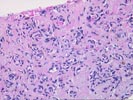
Contributed by Randall G. Lee, M.D.
PATIENT HISTORY:
Per referral report, the patient is an elderly female with multiple lesion in liver by CT. R/O metastasis. No known primary. Review of outside material.


Final Diagnosis (Case 90)

LIVER, NEEDLE BIOPSY -
  1. BENIGN FIBROMYXOID VASCULAR CONNECTIVE TISSUE, CONSISTENT WITH SCLEROSED HEMANGIOMA.
  2. NO EVIDENCE OF MALIGNANCY.

Previous Biopsies on this Patient:
None

TPIS Related Resources:
Liver Transplant Topics
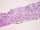

Gross Description - Case 90

The specimen consists of three (3) consult slides and one (1) paraffin block with an accompanying surgical pathology report.


Microscopic Description - Case 90

The liver biopsy contains a small fragment of unremarkable hepatic parenchyma. The bulk of the specimen consists of vascular collagenized tissue with a sparse scattering of inflammatory cells and occasional hemosiderin-laden macrophages. There are numerous blood vessels ranging from slightly dilated venular structures to small capillaries and arterioles. No evidence of malignancy is identified. Overall the changes suggest a sclerosed hemangioma, and this possibility should be correlated with the radiographic and clinical features.


Please mail comments, corrections or suggestions to the TPIS administration at the UPMC.