Previous Biopsies on this Patient:
None
TPIS Related Resources:
Liver
Transplant Topics


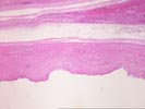
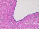
The sections show a multiloculated cyst lined by a flattened to cuboidal, simple epithelial lining. Focally the epithelium shows a suggestion of a brush border or possible cilia, but there is no proliferative lesions or stromal invasion identified. The underlying stroma is predominantly dense fibroconnective tissue with occasional strands of smooth muscle. Deeper in the wall is loose connective tissue containing numerous, irregularly distributed arteries, venous channels and small bile ductules together with small islands of hepatocytes. The ductules are focally grouped in small lobules and are associated with scattered larger ducts. Some of the hepatocyte islands show peripheral ductular metaplasia. This tissue, which is presumably part of the cystic mass lesion, lacks the well-ordered appearance of normal hilar tissue, and suggests a hamartomatous formation.
This lesion does not conform well to any the typical nosologic categories of hepatic cysts. Similar lesions have been reported in the literature as hepatobiliary cystadenomas, although they are unlike the usual such tumor, which almost always occurs in women and has a distinctive mesenchymal stroma. Similarly, the lesion is unlike the usual forgut cysts, which are generally unilocular and have a better developed muscular wall. Because of the haphazard arrangement of biliary and vascular structures in the wall, we would instead favor a hamartomatous process, but are unable to locate any similar reported lesions. This problem of nomenclature is perhaps largely academic, since the lesion is benign and appears to have been completely removed.