Previous Biopsies on this Patient:
None
TPIS Related Resources:
Liver
Transplant Topics


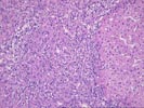
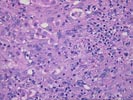
The wedge biopsy of liver consists largely of subcapsular parenchyma, which makes conclusions about the architecture somewhat tenuous. Nonetheless, advanced cirrhosis appears to be present, as manifest by totally deranged architecture with parenchymal nodules bounded by irregular zones of fibrosis. There is mild inflammatory activity in terms of intranodular and interface inflammation. In addition, zones of dense ductular proliferation are prominent. These demonstrate mild nuclear atypia, but overall the changes are reactive in nature. We find such zones in some 5-10% of explanted cirrhotic livers, and often they correspond to areas in which hepatic venules have been occluded and led to parenchymal collapse. No ground-glass hepatocytes, viral inclusions, cytoplasmic globules, iron deposition, bile duct loss, or changes of steatohepatitis are seen.