Previous Biopsies on this Patient:
None
TPIS Related Resources:
Liver
Transplant Topics


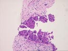
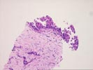
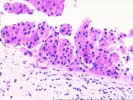
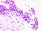
The two cores of liver tissue consist largely of dense fibrous tissue containing scattered mononuclear cells, pigmented laden macrophages and numerous proliferated bile ductules. The fibrosis fades into intact but atrophic hepatic plates with unremarkable liver cells. The proliferated ductules have enlarged hyperchromatic nuclei and an anastomosing pattern; although atypical, their appearance is insufficient for a malignant diagnosis. Of particular additional note, however, are several nodules of dysplastic liver cells located within the fibrosis and well as in separate blood-associated fragments. These nodules show thickened hepatic plates (ranging from four to eight cells in thickness) and demonstrate a trabecular architecture. Individual cells have enlarged, uniformly hyperchromatic nuclei with prominent nucleoli and accentuated nuclear membranes; occasional mitotic figures are also seen. In addition, many cells contain large eosinophilic cytoplasmic inclusions; such inclusions may consist of alpha-fetoprotein or other hepatic-derived proteins and appropriate immunostains and serum AFP determinations may be helpful. Because of the architectural and cytologic abnormalities, these nodules are considered diagnostic of hepatocellular carcinoma. The presence of several discrete, scattered nodules within the specimen suggests a multifocal, diffusely infiltrating pattern of tumor growth within the liver. In addition, in the fibrous stroma are foci of large bizarre cells with cytoplasmic eosinophilia and marked nuclear atypia; these likely represent areas of stromal invasion by the hepatocellular carcinoma.
Thank you for sending this challenging case. We would appreciate any follow-up information you receive.