Comment:
The liver biopsy shows a combination of hepatitic and biliary
changes and thus poses a difficult diagnostic problem. Such a
pattern can be seen in some cases of primary biliary cirrhosis
(which would also account for the granulomas), autoimmune
cholangitis, or poorly defined chronic hepatitic/cholangitic
overlap syndromes. Primary sclerosing cholangitis is a
possibility, but characteristic histologic features are not
present and the overall pattern is somewhat unusual for that
diagnosis. Cholangiography may be required to exclude this
possibility.
Previous Biopsies on this Patient:
None
TPIS Related Resources:
Liver Transplant Topics


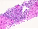
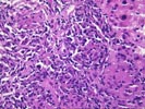
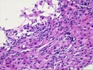
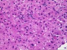
The liver biopsy shows normal lobular landmarks, but the portal tracts are expanded and fibrotic. Most of the portal tracts contain a dense mononuclear infiltrate compose of lymphocytes with occasional plasma cells. This is associated with mild piecemeal necrosis. In addition, the portal tracts contain a granulomatous inflammatory component which is composed of loose accumulations of epithelioid cells in one portal tract and inclusion-bearing giant cells in two others. Bile duct loss is not evident, and classic florid duct lesion are not seen. Several portal tracts show a prominent lymphocytic cholangitis with bile duct injury and infiltrating lymphocytes, whereas other portal tracts demonstrate portal edema and ductular proliferation together with swelling and luceny of periportal liver cells suggestive of chronic cholestasis. The lobules show minor reactive changes.
Because of the granulomatous component, microbial stains should be obtained although the yield is apt to be low. Sarcoidosis is another unlikely consideration. Because of the granulomas and the prominent inflammatory component of the portal process, primary biliary cirrhosis or autoimmune cholangitis would be leading choices, although the absence of more demonstrable bile duct loss argues somewhat against this. An unusual "hepatitic- form" of primary sclerosing cholangitis can not be excluded and may require bile duct imaging to confirm. Please send any follow up information you receive.