Comment:
The immunocytochemical stains are consistent with this
interpretation.The case has also been reviewed by Dr. N. Paul Ohori who agrees
with this interpretation.
Previous Biopsies on this Patient:
None
TPIS Related Resources:
Liver
Transplant Topics

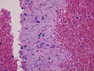
The specimen consists of two portions of fine needle aspiration biopsy. Both are characterized by extensive hemorrhage and blood elements with scanty epithelial and stromal cells. In one slide (labeled 1A) a small nest of epithelial cells with eosinophilic cytoplasm, round to irregular nuclei, and moderately prominent nucleoli are seen. The cells resemble hepatocytes. Elsewhere is seen stromal elements and occasional smaller cells resembling biliary epithelium. The special stains are performed and the results are as follows:
Alpha-feto-protein - negative
Factor 13 - +/-
Lewis X antigen -
negative
Leu M1 - positive
B72.3 - negative
Alpha-1-antitrypsin - heavy
background stain
Polyclonal CEA - +/-