PART 2:
ALLOGRAFT LIVER, NEEDLE BIOPSY (Part 2) -
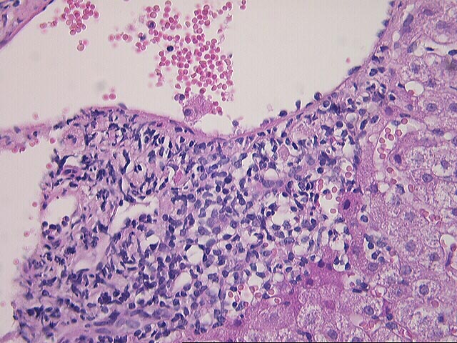
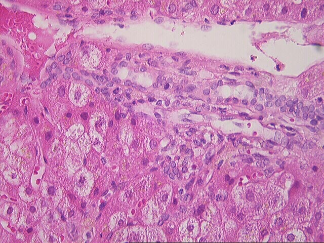
- MILD LOBULAR DISARRAY, HEPATOCYTE SWELLING AND SPOTTY ACIDOPHILIC HEPATOCYTE NECROSIS(see microscopic description).
- MILD CENTRILOBULAR PIGMENT LADENED MACROPHAGE DEPOSITION.
Previous Biopsies on this Patient:
None
TPIS Related Resources:
Liver
Transplant Topics
Gross Description (Case 75)
The specimen consists of nine (9) consult slides with an accompanying surgical pathology report.
Microscopic Description (Case 75)


Part 1:
(1 HE, 1 PAS, 1 Reticulin, 1 CAM5.2, 2 Blanks)
The normal
lobular architecture is distorted because of mild portal expansion, because of
mild mononuclear inflammation and mild cholangiolar reactivity. No bile duct
damage, nor bile duct loss nor portal venulitis is seen. Within most of the
portal tracts, clusters of pigment-laden macrophage deposition are
appreciated. The presence of intact bile ducts is confirmed by CAM5.2
staining.
Throughout the lobule, there is mild disarray with moderate spotty acidophilic necrosis of hepatocytes, sinusoidal lymphocytosis and occasional ceroid-ladened macrophages. No ground glass cells or viral inclusions are seen. No large PAS/D- positive globules are present. The reticulin stain confirms the mildly distorted architecture.
Overall, the biopsy has the histopathological appearance of chronic hepatitis with low-grade activity, consistent with recurrent viral type C. Compared to the previous biopsy (Part 2), the hepatitis seems to be more advanced because of the appearance in the recent biopsy of chronic portal inflammation, without duct damage or venulitis. Although there is no significant rejection activity at this time, clinical follow up with liver injury tests (particularly gamma glutamyltranspeptidase and ALT) and possibly re-biopsy, if indicated, are suggested. The Knodell score is assessed as follows:
| KNODELL'S HISTOLOGY ACTIVITY INDEX | ||
|---|---|---|
| Feature | RANGE | SCORE |
| Periportal and bridging necrosis | (0-10) | 1 |
| Intralobular degeneration and necrosis | (0-4) | 1 |
| Portal inflammation | (0-4) | 3 |
| Fibrosis | (0-4) | 1 |
| TOTAL | (0-22) | 6/22 |
The numerical scoring system of histologic activity index (HAI) has been developed to grade the liver biopsies of chronic active hepatitis. This is based on four categories of periportal and bridging necrosis, intralobular degeneration and necrosis, portal inflammation and fibrosis, with total score of up to 22. This scoring system is correlated well with the severity of disease. A copy of the original paper published by the American Association for the Study of Liver Disease [Hepatology 1:431, 1981] is available at the Department of Pathology upon request.
The numerical scoring system of histologic activity index (HAI) has been developed to grade the liver biopsies of chronic active hepatitis. This is based on four categories of periportal and bridging necrosis, intralobular degeneration and necrosis, portal inflammation and fibrosis, with total score of up to 22. This scoring system is correlated well with the severity of disease. A copy of the original paper published by the American Association for the Study of Liver Disease [Hepatology 1:431 (1981)] is available at the Department of Pathology upon request.
Part 2:
(1 HE)
The normal liver architecture is intact. Eight portal
tracts are identified, all of which are free of inflammation. Bile duct are
present in normal numbers and are intact.
Throughout the lobule, there is mild spotty acidophilic necrosis of hepatocytes. Marked golden green pigment deposition is seen in centrilobular hepatocytes and macrophages. Diffuse hepatocellular swelling is also appreciated.
Overall, the histopathological changes are not specific for a particular etiology, but are suggestive of early recurrent type C viral hepatitis. Azathioprine treatment might also have contributed to the changes, because withdrawal of the agent apparently resulted in improvement of the liver injury test results.
Please mail comments, corrections or suggestions to the TPIS administration at the UPMC.
