Comment:
The loss of bile ducts corresponds to the clinical
picture of cholestasis and suggests a ductopenic syndrome. The most likely
possibilities, given the histologic appearances, are drug-induced or
idiopathic ductopenia. Primary biliary cirrhosis, autoimmune cholangitis, and
primary sclerosing cholangitis are much less likely possibilities.
Previous Biopsies on this Patient:
None
TPIS Related Resources:
Liver
Transplant Topics


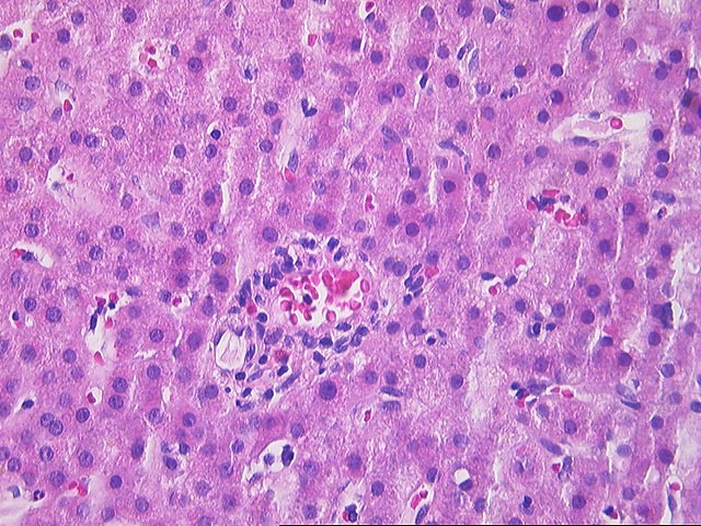
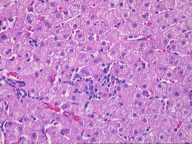
The liver biopsy shows overall intact hepatic architecture although portal tracts are small and often difficult to identify. In addition, several of the recognizable portal tracts lack an intralobular bile duct and instead contain a sparse infiltrate composed of a few mononuclear cells with scattered neutrophils and eosinophils. In occasional portal tracts the bile duct shows mild epithelial reactivity. In addition, the lobules contain a sparse but widespread sinusoidal infiltrate of mononuclear cells but this is associated with only minor liver cell injury. Mild focal macrovesicular steatosis is identified.
Overall, the changes suggest a ductopenic syndrome. Although they can not be completely excluded, primary biliary cirrhosis and primary sclerosing cholangitis are unlikely possibilities. There is no evidence for sarcoidosis or some of the other unusual causes of ductopenia. The remaining diagnostic considerations would include drug-induced or idiopathic forms. The patient is apparently not on any of the handful of drugs implicated in ductopenia. Idiopathic ductopenia, as recently reviewed (NEJM 1997; 336:835), often occurs in asymptomatic, middle-aged woman and infrequently leads to progressive liver disease.