Previous Biopsies on this Patient:
None
TPIS Related Resources:
Liver Transplant Topics


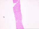
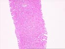
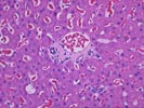
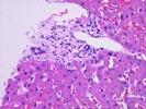
(2 HE), The normal lobular architecture is intact and approximately seven small portal tracts are identified, two of which are devoid of ducts. Several of the other of the triads show mild cholangiolar reactivity and occasional eosinophils. An occasional lymphocyte is seen in the bile duct epithelium but overall, the ductal degeneration are not particularly striking. There is no interface activity and overall the portal inflammation is mild.
Throughout the lobules, there is mild Kupffer's cell hypertrophy but no evidence of lobular disarray. Mild panlobular sinusoidal red blood cell congestion is seen. There is also focal thickening of the plates and focal sinusoidal dilatation. No ground glass cells, nor prominent pigment deposition, nor viral inclusions are seen.
Overall, the histopathologically changes are relatively mild and non-specific. The differential diagnosis of an elevated alkaline phosphatase (as per the clinical history) in a liver allograft recipient includes cholestatic disorders such as biliary tract obstruction or stricturing, chronic rejection, ischemic injury, cholestatic drug or viral hepatitis and nodular regenerative hyperplasia. Unfortunately, the histopathological changes are insufficient for a definitive diagnosis. Of the possibilities listed above, hepatitis and chronic rejection can probably be excluded. This leaves a low grade nodular regenerative hyperplasia-type changes or low grade biliary tract obstruction or stricturing. If the liver injury tests remain elevated, continued follow-up will probably be needed to determine the underlying cause.