Previous Biopsies on this Patient:
None
TPIS Related Resources:
National Cancer Institute PDQ treatment information on liver cancer
Liver Transplant Topics

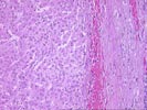
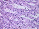
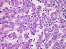
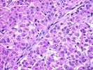
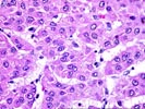
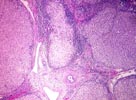
All of the sections demonstrate similar features. There is a well-differentiated hepatocellular carcinoma characterized by hepatocytes with generally mild nuclear atypia variably arranged in trabecular and pseudoglandular architectural patterns with cellular crowding and hepatic plates of more than three cells thickness. The tumor is bounded by a thick fibrous pseudocapsule, which is focally invaded, and no bile ductules are noted along the periphery of the tumor nests. The tumor contains numerous aberrant arteries, scattered foci of inflammatory cells, and several areas of greater degree of nuclear atypia and pleomorphism. No definite vascular invasion is identified. Although the tumor presented with hemoperitoneum and presumably invaded through the liver capsule, this is not clearly documented histologically. The background liver demonstrates a well developed cirrhosis, mixed macronodular and micronodular type, with mild inflammatory activity. No viral inclusions, ground glass hepatocytes, pigment deposition, cytoplasmic globules or evidence of steatohepatitis is seen. No particular etiology is suggested, although the overall pattern and presence of occasional lymphoid aggregates raises the possibility of chronic hepatitis C, and this should be excluded with appropriate serologic tests.
Although in areas the tumor is very well-differentiated and may, in isolation, be difficult to distinguish from an atypical macroregerative nodule (dysplastic nodule), the overall features are indicative of a hepatocellular carcinoma. The crucial findings include the well-formed trabecular architecture with cellular crowding and thickened cell plates, the pseudoglandular formation in more than an isolated focus, the absence of peripheral bile ductules, the abundance of aberrant arteries, and the foci of overt nuclear atypia.