Previous Biopsies on this Patient:
None
TPIS Related Resources:
National Cancer Institute PDQ treatment information on liver cancer
Liver Transplant Topics
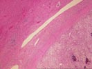
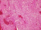
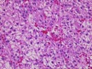
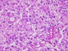
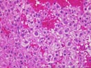
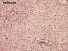
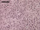
(5 H&E, 1 Reticulin) Sections from the liver show an intact lobular architecture with mild intralobular regenerative changes. Near the grossly identified mass, portal changes, typical of a mass effect, are seen.
The most significant finding is the presence of a large hepatocellular neoplasm. It appears to be largely surrounded by a fibrous capsule and/or fibrous tissue. No definite vascular invasion is seen. However, the lesion is highly cellular and contains multiple "aberrant" arteries. It is composed of small hepatocytes and abundant bile production with pseudoacinar formation can be seen. Cytologically, many of the small hepatocytes comprising the lesion are quite small and some contain eosinophilic globules. For the most part, they also contain small a small round nucleus and a small amount of eosinophilic cytoplasm. The N:C ratio is increased in some areas throughout the tumor. In addition, focal clear change is seen.
On closer examination, an increased mitotic rate is seen in some areas up to 1/2-3 HPF. Individual apoptotic tumor cells are also seen.
The reticulin stain nicely demonstrates focal paucity of reticulin fibers in between individual clusters and sheets of the neoplastic cells. This is in contrast to the intact reticulin architecture in the surrounding liver.
Overall, I feel these findings are indicative of a well- differentiated hepatocellular carcinoma. This contention is based on the age and sex of the patient and the histopathological findings. The histologic findings in favor of a malignant diagnosis include the cellularity of the lesion, the increased mitotic rate, and focal attenuation of the reticulin architecture. I have taken the liberty of sharing this case with Drs. Nalesnik and Lee, who also concur with this diagnosis.