Comment:
Given its histologic appearance and the results of an
immunohistochemical panel, this carcinoma is most likely of
metastatic origin, although cholangiocarcinoma is also a
possibility. No definitive evidence of hepatocellular
differentiation is demonstrated.
Previous Biopsies on this Patient:
None
TPIS Related Resources:
National Cancer Institute PDQ treatment information on liver cancer
Liver Transplant Topics
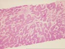
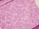
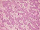
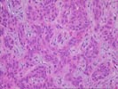
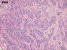
The liver biopsy specimen contains several areas of poorly differentiated carcinoma characterized by anastomosing cords of overtly malignant cells. These are separated by interdigitating loose desmoplastic stroma with scattered mixed inflammatory cells. The individual tumor cells have moderate amphophilic cytoplasm and large pleomorphic nuclei with soft dispersed chromatin and frequent bizarre mitoses. No bile production is identified. There is focal tumor cell apoptosis in addition to more extensive confluent necrosis within the tumor. The tumor abuts relatively normal background liver that shows portal tract expansion and edema with ductular proliferation and an associated inflammatory population, changes consistent with obstructive type changes occurring in the vicinity of mass lesions.
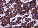
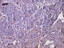
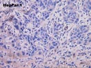
To better characterize the nature of the tumor, a number of stains were performed, both at the referring institution and here. The PAS stain is negative for mucin production. CAM 5.2 cytokeratin immunostain is strongly positive, and 34BE12 cytokeratin immunostain appears to be negative, although the stain is difficult to interpret because the bile ducts in the background liver, which should be be positive, are not staining. The alpha fetoprotein immunostain shows focal positivity, but again the interpretation is complicated because similar positivity is noted in the normal hepatocytes. The monoclonal CEA immunostain is negative. In addition, a polyclonal CEA immunostain was performed which fails to show either cytoplasmic or canalicular reactivity, and a HepPar 1 stain is likewise negative. This combination of reactivity does not provide strong evidence for a specific line of differentiation, and specifically does not support hepatocellular differentiation. For this reason we favor the carcinoma representing either a metastatic carcinoma from an extrahepatic site or possibly a cholangiocarcinoma.
Thank you for sending this challenging case and I would appreciate any further information you receive.