Comment:
The biopsy most likely represents subcapsular tissue. The
overall process is suggestive of a cirrhotic pattern compatible
with hepatitis C infection, consistent with the clinical history.
However, the change may be focal and non-representative of the
liver as a whole. Given the degree of fibroinflammatory change
in this biopsy, comment regarding the presence or absence of
rejection elsewhere is not possible. The inflammation, although
dense, does not appear to have the atypia associated with post
transplant lymphoproliferative disease.
Previous Biopsies on this Patient:
None
TPIS Related Resources:
Liver Transplant Topics


(8 HE)
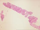
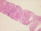
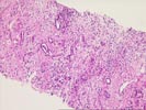
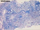
The biopsy consists of a fragmented core of tissue with
recognizable bile ducts and ductules, and rare hepatocytes. On
one end of the specimen appears dense regular connective tissue,
possibly capsular. Vessel and duct proliferation is noted
throughout the remainder of the specimen, together with a
moderately dense predominantly chronic inflammatory infiltrate.
No atypical cells or mass lesions are appreciated. Bile ducts
occasionally show slight disorientation of nuclear placement, but
in general appear unremarkable.