Per referral letter, the patient is a 49-year-old female with a large, vascular neoplasm in her liver. On May 5th, the patient was taken to the operating room in the hope of removing the lesion through a left lobectomy. Upon opening the abdomen, the surgeon discovered that there were at least eight cherry-red lesions in the right lobe that blanched when he pressed on them and promptly refilled when he released them. The surgeon deemed the patient unresectable and took a wedge biopsy of one of these lesions. Review of outside material.
Comment:
This tumor is not typical of any of the usual variants of hepatic
vascular malignancies. Overall it is probably best considered as
a low-grade angiosarcoma, but we are not certain that its
biologic behavior will be that of the usual hepatic angiosarcoma.
Although certain features (including the patient's age and sex)
prompted consideration of an epithelioid hemangioendothelioma,
the histologic findings are not entirely compatible with that
diagnosis.
Previous Biopsies on this Patient:
None
TPIS Related Resources:
National Cancer Institute PDQ treatment information on liver cancer
Liver Transplant Topics
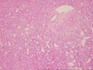
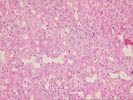
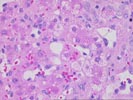
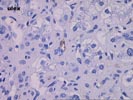
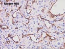
The wedge biopsy of liver shows an ill-defined infiltrative mass composed of scattered atypical cells contained within zones of atrophic hepatic plates, fibrosis, and ductular proliferation. These cells also extend into the parenchyma, where they line the sinusoids, which are frequently irregularly dilated. Focal tufting of cells is noted in larger vein branches. Individual cells display large, hyperchromatic nuclei, and occasional bizarre mitotic figures are seen. The cells are of endothelial origin, as demonstrated by reactivity for Ulex europeus, Factor VII antigen, and a CD34 stain performed here. Immunostains for factor XIIIA show a few positive cells in the more solid regions.
This tumor, although clearly a malignant vascular neoplasm, poses a problem in assigning a diagnostic category. We have designated it a low-grade angiosarcoma, but would not assume that it will demonstrate the omnious natural history of the usual hepatic angiosarcoma and, in particular, would not exclude liver transplantation if the patient is otherwise a reasonable candidate.