Previous Biopsies on this Patient:
None
TPIS Related Resources:
National Cancer Institute PDQ treatment information on liver cancer
Liver Transplant Topics


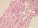
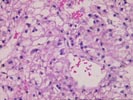
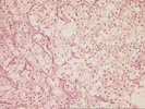
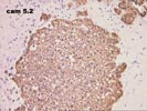
The liver biopsy demonstrates several fragments of hepatocellular carcinoma characterized by irregular trabeculae of well differentiated malignant hepatocytes with focal clear cell differentiation. Several fragments contain dense fibrosis containing hemosiderin-laden macrophages. The tumor abruptly abuts the adjacent fibrosis without any intervening ductular reaction. The reticulin stain demonstrates focal thickening of the cell plates with patchy loss of reticulin, consistent with a hepatocellular carcinoma. Immunostains show positive reaction for CAM5.2 cytokeratin, but negative for alpha-fetoprotein, AE1 cytokeratins, and monoclonal CEA, a pattern of reactivity again consistent with hepatocellular carcinoma. No background liver is present in the sections and thus no assessment of the presence of underlying cirrhosis can be made.