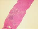
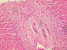
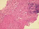
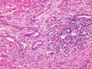
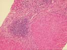
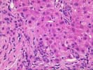
PART 2: LIVER, NEEDLE BIOPSY (2/26/97) -
COMMENT:
The hepatic changes are those of a chronic biliary process
leading to cirrhosis. Because of the absence of either bile duct
loss or obliterative periductal fibrosis, the appearances are
more suggestive of secondary biliary cirrhosis rather than
primary sclerosing cholangitis. Clinical and radiographic
information are needed, however, to confirm this diagnosis.
Primary biliary cirrhosis is another consideration, although
given the histologic changes, it is somewhat less favored.
Previous Biopsies on this Patient:
NONE
TPIS Related Resources:
Knodell Scoring
Liver Transplant Topics
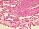
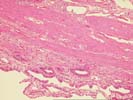
The gallbladder shows mild thickening of the wall by edema and mild fibrosis. The epithelium shows focal reactive changes with edema and occasional inflammatory cells within the lamina propria. Although the changes are mild, they are consistent with a resolving cholecystitis.
The liver biopsy demonstrates portal-based bridging fibrosis with early cirrhosis development. There is a mild patchy mononuclear infiltrate within the lamina propria, but intralobular bile ducts are intact. There is pronounced ductular proliferation with accompanying neutrophils along the periphery of many of the fibrous bands. No granulomas or florid duct lesions are identified. The changes are those of a progressive biliary fibrosis with cirrhosis. Because of the ductular proliferation and absence of bile duct loss, secondary biliary cirrhosis is favored, but primary sclerosing cholangitis and primary biliary cirrhosis should be excluded by appropriate radiographic and imaging studies. A copper stain will be performed to confirm the biliary nature of the process.