PART 2: LIVER, HEPATIC SEGMENT 4, RESECTION -
PART 3: LIVER, WEDGE BIOPSY -
Previous Biopsies on this Patient:
NONE
TPIS Related Resources:
Knodell Scoring
Liver Transplant Topics


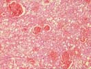
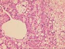
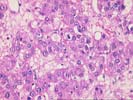
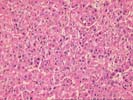
The tumor consists of hepatocytic cells arranged generally in thickened cords with occasional foci of cellular crowding and trabecular formation and prominent areas of hemorrhage, pelotic- like change, and necrosis with organization. The tumor cells contain pronounced steatosis and have variably acidophilic and cleared cytoplasm. There is a mild degree of cytologic atypia characterized by nuclear enlargement, hyperchromasia, and crowding with minor pleomorphism. Mitoses are rare, and no vascular invasion is identified. The differential diagnosis includes hepatocellular adenoma and hepatocellular carcinoma, but based on the mild cytologic and architectural atypia and the clinical setting, we favor a diagnosis of well differentiated hepatocellular carcinoma. The separately submitted liver biopsy demonstrates patchy mononuclear portal infiltrate with mild portal fibrosis with minimal piecemeal necrosis and foci of large cell change in the background. No viral inclusions, ground glass hepatocytes, pigment deposition, cytoplasmic globules, or features of cirrhosis are identified. The possibility of chronic hepatitis cannot be excluded, and should be explored on the basis of clinical findings and laboratory tests.
This is a difficult case, but in our experience, tumors with this mild degree of atypia can recur or metastasize. Although this case was sent to Dr. Demetris, he was not available when it was received.