COMMENT:
Although the routine hematoxylin and eosin appearance of the
tumor suggests a metastatic carcinoma, the panel of
immunocytochemical stains is most consistent with hepatocellular
origin of the tumor. It is possible that this may represent an
intrahepatic metastasis. The case has also be reviewed by Dr. Randall
Lee who agrees with the interpretation of the special stains.
Clinical correlation is requested.
Previous Biopsies on this Patient:
NONE
TPIS Related Resources:
National Cancer Institute PDQ treatment information on liver cancer
Knodell Scoring
Liver Transplant Topics
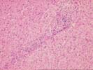
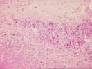
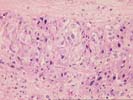
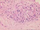
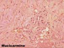
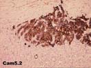
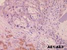
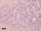
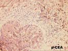
There is a core of hepatic parenchyma which shows portal inflammation of a mild to moderate degree. In addition there are several smaller fragments which also contain hepatocytes but additionally contain necrotic areas and areas of anaplastic epithelial cells. The cells assume an elliptical architecture, suggesting the possibility that they may be within vascular or lymphatic spaces, at least in areas. The cells have an amphophilic to eosinophilic cytoplasm with a nuclear to cytoplasmic ratio of approximately 1:1. Nuclear membranes are thickened, rounded and slightly irregular. Nucleoli are prominent, single and central. The special stains are as follows: alphafetoprotein, negative x 2; polyclonal CEA, canalicular pattern; monoclonal CEA, negative; CAM5.2, positive with membrane accentuation; AE1, negative; mucicarmine equivocal to negative; HEPPAR-1, negative; synaptophysin, negative; S-100, negative; alpha-1-antichymotrypsin, negative; AE1/AE3, positive. There is mild mononuclear inflammation within the lobules as well as within the periportal regions. Generalized hepatocellular swelling is seen but frank necrosis is rare. The HAI score is as follows:
| KNODELL'S HISTOLOGY ACTIVITY INDEX | ||
|---|---|---|
| Feature | RANGE | SCORE |
| Periportal and bridging necrosis | (0-10) | 3 |
| Intralobular degeneration and necrosis | (0-4) | 3 |
| Portal inflammation | (0-4) | 3 |
| Fibrosis | (0-4) | 3 |
| TOTAL | (0-22) | 12 |
The numerical scoring system of histological activity index (HAI) has been developed to grade the liver biopsies of chronic active hepatitis. This is based on four categories of periportal and bridging necrosis, intralobular degeneration and necrosis, portal inflammation and fibrosis, with total score of up to 22. This scoring system is correlated well with the severity of disease. A copy of the original paper published by American Association for the Study of Liver Disease [Hepatology 1:431 (1981)] is available at the Department of Pathology upon request.