PART 2: APPENDIX, RESECTION -
COMMENT:
As always, a histopathological diagnosis of cirrhosis should be
clinically confirmed by searching for portal hypertension and
other stigmata of chronic liver disease. This is especially true
in the subcapsular region, where the fibrosis may be more
advanced than in the deeper parenchyma.
Previous Biopsies on this Patient:
NONE
TPIS Related Resources:
Knodell Scoring
Liver Transplant Topics
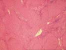

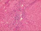
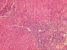
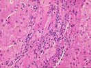
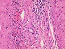
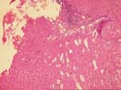
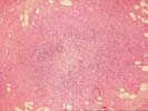
The wedge biopsy of liver is from the subcapsular region. The normal lobular architecture is largely replaced by an active, predominantly macronodular cirrhosis. The normal spatial relationship between portal tracts and central veins has been completely disrupted in most areas. In addition, several nodules, surrounded on all four sides by fibrosis, are seen.
On closer examination, there is a mild to moderate mononuclear inflammatory cell infiltrate within the fibrous septae. Bile ducts are intact, and no florid duct lesions are seen. Interface activity is appreciated. Throughout the lobules/nodules, there is Kupffer cell hypertrophy and occasional acidophilic bodies. Focal oncocytic metaplasia of the hepatocytes and cautery artefact are also seen. No definite ground glass cells are identified, and no prominent increase in pigment is seen.
Overall, the changes are indicative of significant chronic inflammatory liver disease, such as chronic Hepatitis B or C virus infection. Clinical correlation is suggested.
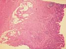
An additional section is submitted, which is apparently from the appendix. It shows focal fibrosis and mild chronic inflammation. Fibrous obliteration of the tip is also present.