 Contributed by Michael Nalesnik, M.D.
PATIENT HISTORY:
The patient is an elderly man with a clinical history of HH, severe duodenitis, inflammatory polyp of duodenum.
Final Diagnosis (Case 5)
PART 1:DUODENUM, ENDOSCOPIC BIOPSY -
- TUBULOVILLOUS ADENOMA, DUODENUM (see comment).
PART 2:GASTROESOPHAGEAL JUNCTION, ENDOSCOPIC BIOPSY -
- BARRETT' S ESOPHAGUS WITH LOW GRADE DYSPLASIA.
- ACUTE AND CHRONIC INFLAMMATION, GASTROESOPHAGEAL JUNCTION, CONSISTENT WITH REFLUX ESOPHAGITIS.
Microscopic Description - Case 5
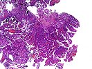
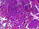
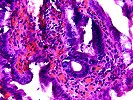
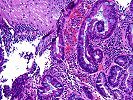
Slide labeled A consists of a portion of small bowel mucosa with glands consistent with Brunner' s glands. The mucosal epithelium shows a villous configuration in some areas with variable congestion noted. The epithelium ranges from unremarkable in appearance to that of a more crowded aggregate of mucosal cells. The latter have an increased nuclear to cytoplasmic ratios, slightly hyperchromatic, but smooth contoured nuclei and moderately prominent nucleoli. Some irregularity in luminal shape is noted, consistent with tortuous glands. No invasion into the stroma is seen. In some regions this epithelium is associated with acute inflammation, whereas in others an acute inflammatory component is not seen. The underlying glands appear unremarkable. No evidence of viral inclusions is appreciated.
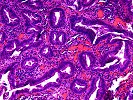
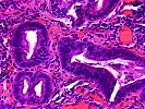
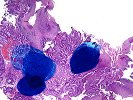
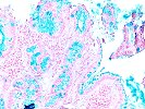
Slide labeled B1 consists of a combination of squamous mucosa and mucous epithelial cells. The former shows normal maturation with increased numbers of acute and chronic inflammatory cells and no evidence of dysplasia. The mucosal epithelial cells are remarkable for numerous goblet cells and focal irregularity and crowding of individual glands. In one area, previously noted and marked with ink, a small aggregate of crowded glands show increased nuclear to cytoplasmic ratios in relation to other glands, as well as occasional goblet cell dysplasia. Scattered mitoses are noted. No invasion of underlying stroma is seen. No high grade dysplasia or carcinoma in-situ is appreciated at this time. Elsewhere, a combination of goblet cells and columnar absorptive type cells line the surface. Scattered Paneth cells are noted as well. An alcian blue stain is strongly positive both in goblet cells and in the mucin overlying the absorptive epithelial cells.
Previous Biopsies on this Patient:
None
TPIS Related Resources:
None.
Gross Description - Case 5
Received for consultation are a total of three (3) consult slides.
Photomicrographs - Case 5
Please mail comments, corrections or suggestions to the
TPIS administration at the UPMC.
)
)
)
)
)
)
)
)