PARTS 3 AND 4:
STOMACH, ANTRUM AND CARDIA, ENDOSCOPIC BIOPSY
-
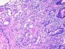
- MILD CHRONIC ACTIVE GASTRITIS WITH HELICOBACTER ORGANISMS
IDENTIFIED.
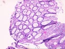
- CAUTERIZED COLONIC MUCOSA SHOWING FEATURES OF HYPERPLASTIC POLYPS.
- NO EVIDENCE OF ADENOMA OR MALIGNANCY.
PART 5:
ESOPHAGUS, ENDOSCOPIC BIOPSY -
- MILD ACTIVE ESOPHAGITIS CONSISTENT WITH REFLUX DISEASE.
PARTS 6 AND 7:
LARGE BOWEL, COLON AT 40.0 CM. AND RECTUM, ENDOSCOPIC
BIOPSIES -
Comment:
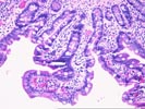
The duodenal biopsies show evidence of previous
mucosal injury with regenerative changes. These changes are not felt to be
dysplastic or adenomatous in nature based on the lack of increase in
glandular profiles, the concomitant presence of active inflammatory
infiltrate, and the maturation towards the luminal surface. These findings
are all in favor of a reactive rather than neoplastic proliferation.
Previous Biopsies on this Patient:
None
Gross Description - Case 4
The specimen consists of eight (8) consult slides with an accompanying surgical pathology report.Microscopic Description - Case 4


(8 HE)
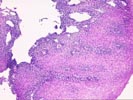
Microscopic examination substantiates the above diagnosis.
Please mail comments, corrections or suggestions to the TPIS administration at the UPMC.