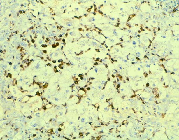
Figure 6.
This stain decorates mature tissue and infiltrative macrophages, which are
numerous and appear reactive in fibrosing cholestatic hepatitis. Note the
positive staining cells in the sinusoids, surrounding clusters of swollen and
ballooned hepatocytes. These changes are typical of fibrosing cholestatic
hepatitis seen with either recurrent or de novo type B viral hepatitis.
Please mail comments, corrections or suggestions to the
TPIS administration at the UPMC.