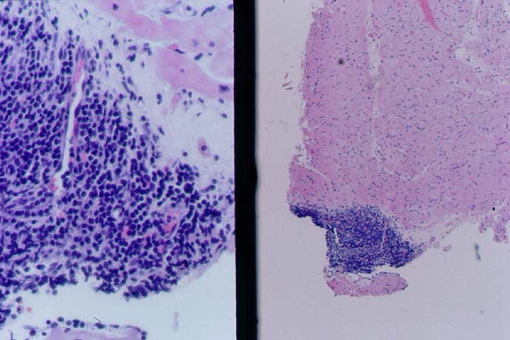
Figure 3.
This is another example of a type A Quilty lesion, which is confined to the
endocardium. The low power view on the right shows the sharp border with the
underlying myocardium. The left side of this photomicrograph illustrates
the small vascular channels.
Please mail comments, corrections or suggestions to the
TPIS
administration at the UPMC.